


INTRODUCTION OF DLC
Leukocytes or white blood cells (WBCs) are blood cells which form an integral part of the immune system of the body. There are five types of WBCs with each of them having different functions. Differential Leukocyte Count Test measures the total number of all the WBCs in blood.
Type of Leucocytes.
Lymphocytes.:- There are two main types of lymphocytes: B cells and T cells. B cells fight off invading viruses, bacteria, or toxins. T cells target and destroy the body's own cells that have been infected by viruses or cancer cells.
Monocytes r:-foreign material, remove dead cells, and boost the body's immune response.
Eosinophils:- fight infection, inflammation, and allergic reactions. They also defend the body against parasites and bacteria.
Basophils:- release enzymes to help control allergic reactions and asthma attacks.

IMPORTANCE OF DLC.
The Differential Leukocyte Count Test is performed:
- As a part of Complete Blood Count (CBC) Test in regular health checkups.
- As follow up test in case of abnormal CBC results.
- To help diagnose infections and inflammation.
- To help diagnose conditions that decrease WBC count like bone marrow disorders.
- To monitor bone marrow function.
- To monitor chemotherapy treatment.
REQUIREMENTS FOR DLC.
- Leishman stain.
- Microscopic glass slide.
- Distill water.
- Blood specimen.
- Spreader.
- Manual Cell counter.

PROCEDURE.
- Prepare a thin blood smear on a clean and dry microscopic glass slide.
- Air dry it.
- Now cover the whole dried thin blood smear with undiluted leishman stain solution by counting the drops of leishman stain.
- After two minutes add the twice amount of distilled water and mix the content by swirling and wait for 8 minutes.
- Rinse the slide.
- Air dry the slide in a tilted position so that the water easily remove out of the slide.
- Observe the tail to head position of slide in zig-zag movement under oil immersion objective lens of the microscope.
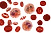
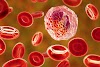
OBSERVATION
- Neutrophil :- purple colored nuclei with pick cytoplasm.
- Eosinophil :- cytoplasm is faint pink, nucleus is purple and granules are orange red.
- Basophils :- granules is dark blue with purple nucleus.
- Monocytes :- pink cytoplasm with purple color nucleus.
- Lymphocytes :- Dark blue nucleus with light blue cytoplasm.

NORMAL RANGE.
- Neutrophil = 40-70%
- Eosinophil = 1-6%
- Basophils = 0-1%
- Monocytes =2-8%
- Lymphocytes = 20-40%
CLINICAL SIGNIFICANCE


.)
0 Comments